
Synthesis of ABO antigens, like all carbohydrate antigens, is the result of a complex series of interactions between a range of different glycosyltransferases residing in the endoplasmic reticulum and Golgi apparatusColley, 1997Maccioni et al.,2011aMaccioni et al., 2011b. To form an antigen these glycosyltransferases must catalyse reactions in an orderly manner between a variety precursors and substrates in an intracellular organelle environment where appropriate organization, transport, and assembly are also required. This environment is competitive, co-operative and dynamic, and fragile where even subtle changes in variables and environment may substantially influence not only the amount of resultant antigen but also its final structure.
Blood group A and B antigens are synthesized by the glycosyltransferase enzyme N-acetylgalatosaminyltransferase and galactosaminyltransferase which catalyzes the transfer of GalNAc (using UDP-GalNAc) and Gal (using UDP-Gal), respectively, to the OH-3 position of the terminal Gal of the H structure (Fucα2Gal) to create the A and B antigens Palcic et al., 2011Watkins & Morgan, 1959Tuppy & Staudenbauer, 1966. Manganese ions (Mn2+) are required as a co-factors and the H disaccharide Fucα1-2Gal residue is the minimal required acceptorClausen & Hakomori, 1989Morgan& Watkins, 2000.
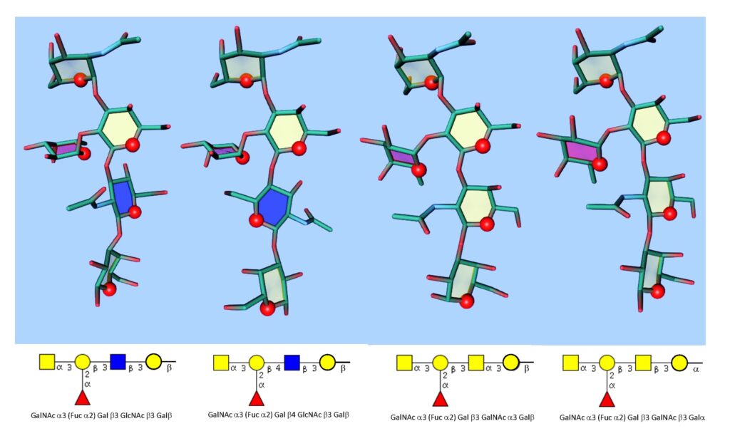
Although the minimal structures representing the A and B antigens are clearly defined, these trisaccharide determinants are always present on a variety of peripheral disaccharide cores (Table 1).
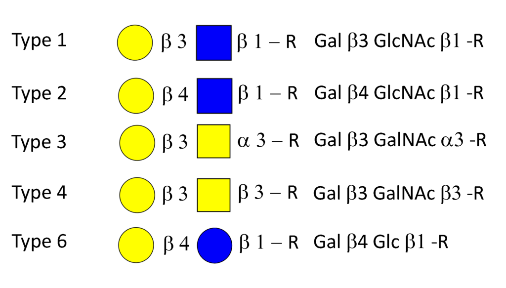
Each of which imparts specific 3D antigenic features (Figure 4).
The most frequent antigens found in red cells are carried by a type 2 chain Rege et al., 1963. Other types of antigens described on red cells are carried by type 3 and type 4 chains Donald, 1981Clausen et al., 1986aClausen et al., 1986bBremer et al., 1984. Evidence for blood group A with a type 6 peripheral core has also been reported on red cells Holgersson et al., 1992Svensson, et al., 2011. The type 1 antigens as glycolipids are also carried by red cells but these are absorbed from the plasma onto the red cell and may, if present, exist in one of two forms ; for example A type 1 or the compound antigen ALeb depending on whether or not they have been modified by Lewis fucosylation (see below). The ratio, quantity, and elongation of these antigens in the red cell membrane is determined by a complex interplay of the relative levels of activity of the ABO, Secretor and Lewis fucosyltransferases together with environmental factors Henry et al., 1995Mollicone et al., 1995.